Researchers have created a new artificial intelligence system that can spot dangerous blood cells more reliably than human experts, potentially transforming how leukemia and other blood disorders are diagnosed. The technology, called CytoDiffusion, analyzes the shape and structure of blood cells under a microscope with a level of precision that exceeds what trained specialists can achieve.
The system represents a significant step forward in medical diagnostics. Rather than relying on a doctor's visual assessment of blood samples, the AI examines subtle variations in how cells appear that might signal disease. When tested, CytoDiffusion identified abnormal cells linked to leukemia with much higher sensitivity than existing systems, and it performed as well as or better than current leading models even when trained with far fewer examples.
Background
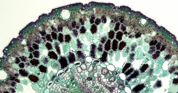
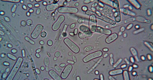
Diagnosing leukemia has always been a challenging task for medical professionals. The traditional process involves a doctor or specialist examining a blood sample under a microscope, looking for distinctive patterns and abnormalities in cell structure. This work is time-consuming, and the results can vary depending on the experience of the person doing the examination. In large laboratories, this process happens thousands of times every day.
The challenge becomes even more difficult with certain types of leukemia. Some forms of the disease display clear, recognizable signs that experienced doctors can spot relatively easily. But other subtypes are far more subtle. In fact, researchers have found cases where even the most experienced cytologists—specialists trained in examining cell structure—cannot detect abnormalities under the microscope that the AI system can identify.
This is where artificial intelligence comes in. The same type of technology used to power image generators like DALL-E has been adapted to analyze blood cells in unprecedented detail. Rather than simply learning to sort cells into fixed categories, the AI models the entire range of how blood cells can appear, making it more resilient to differences between hospitals, microscopes, and staining techniques.
Key Details
CytoDiffusion works by studying patterns in blood cell appearance that humans might miss or not perceive at all. The system was trained by feeding it images of blood samples from patients with leukemia as well as images of healthy blood cells. The artificial neural networks analyze these differences and learn to recognize typical patterns associated with disease.
One of the most important features of CytoDiffusion is that it can quantify how confident it is in its own predictions. This means doctors can see not just what the AI thinks is happening, but also how certain the system is about that assessment. This built-in measure of confidence helps clinicians understand when they should trust the AI's diagnosis and when they might need additional testing or a second opinion.
In testing, the system demonstrated remarkable results. When researchers compared CytoDiffusion to other leading AI models, it performed as well or better, despite being trained on far fewer examples. This efficiency matters in the real world, where gathering large amounts of labeled training data can be expensive and time-consuming.
The technology also shows promise for detecting rare or unusual cases. In one trial, the AI correctly identified a subtype of leukemia in 75 percent of cases where even the most experienced cytologists could not detect any abnormalities under the microscope. This suggests the computer is recognizing features that humans either do not perceive or have previously ignored.
"Rather than focusing only on obvious patterns, it studies subtle variations in how cells look under a microscope. This makes it more resilient to differences between hospitals, microscopes, and staining techniques, while also improving its ability to detect rare or abnormal cells."
What This Means
The implications of this technology extend beyond just faster or more accurate diagnosis. AI systems like CytoDiffusion could help reduce diagnostic delays, particularly in areas where trained hematologists are scarce or where resources are limited. In low-resource settings around the world, access to expert blood cell analysis has always been a challenge. An AI system that can work anywhere with a microscope and a computer could help level that playing field.
The technology also opens doors for detecting other diseases. The methods that work for identifying leukemia could potentially be adapted to spot other blood disorders or even completely different conditions. Some researchers are already exploring how to apply similar techniques to bone marrow samples, expanding the reach of AI-assisted diagnosis.
However, experts are clear that AI will not replace doctors. The diagnosis of cancer remains a medical decision that needs to be made by a human. Physicians and researchers will need to continually improve the algorithms and adapt them to new clinical questions. The goal is to create a partnership between human expertise and artificial intelligence, where the technology handles the pattern recognition and the doctor makes the final call.
The development of CytoDiffusion and similar systems reflects a broader shift in how artificial intelligence is being applied to medicine. Rather than trying to replace human judgment, these tools are designed to support clinicians, catch things they might miss, and help them work more efficiently. As these systems become more sophisticated and more widely adopted, they could significantly improve outcomes for patients with blood cancers and other serious conditions.